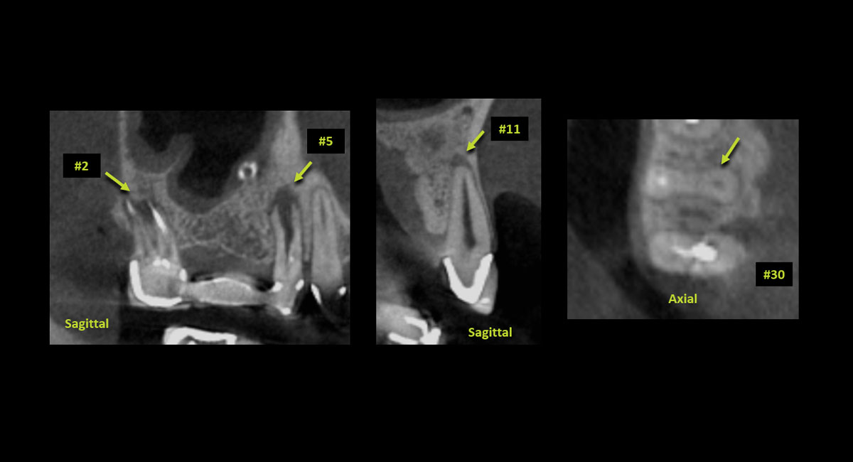
Cone beam computed tomography (CBCT) is a three-dimensional imaging technology that is becoming increasingly popular in endodontics thanks to its ability to provide detailed images of the internal structure of teeth. Several practical benefits for CBCT exist when used before or during root canal treatment.
- CBCT provides an accurate and detailed view of the root and pulpal anatomy.
- CBCT aids in challenging diagnosis and influences the decision for a surgical or non-surgical approach.
- Use of CBCT can lead to more predictable outcomes, improving the efficiency of the practitioner and overall patient satisfaction.
- CBCT Reports written by Oral & Maxillofacial Radiologists ensure proper documentation of all incidental findings as well as the endodontic condition of the teeth in question.
CBCT for Endodontic Diagnosis
The use of CBCT in endodontic diagnosis allows clinicians to evaluate dental anatomy in 3-D, critical for the successful completion of complex cases. Detectors equipped with small pixel sizes (around 0.09 mm) can generate images with high resolution, especially in the absence of gutta percha, endodontic sealer, and high-density restorative materials prone to generate significant artifact.
CBCT for Treatment Planning
CBCT images can reveal the number, size, and shape of canals, as well as any potential obstructions, calcifications, or even root fractures. It can detect root resorption with greater precision than conventional radiographs, especially early invasive cervical resorption lesions. The detailed images produced by CBCT help clinicians determine the appropriate treatment for the tooth in question and identify challenges that may impede successful completion of the procedure. The findings can also guide the selection of materials and orientation of instruments during treatment, leading to more predictable outcomes.
CBCT following Unsuccessful Endodontic Intervention
When patients present with unsuccessful endodontic intervention, CBCT can help identify potential factors such as missed canals or fractures that are preventing full resolution of the initial lesion. If the CBCT suggests treatment success by depicting new bone formation and collapse of periapical lesions, a separate and unrelated condition may have developed. Some examples could include symptomatic adjacent teeth, referred odontogenic or myofascial pain, and acute sinusitis, which must be differentiated from periapical mucositis (when an active endodontic lesion is stimulating sinus floor mucositis).
CBCT for Surgical Endodontic Intervention
In cases where endodontic surgery is necessary, the regional anatomy depicted on CBCT can guide positioning for safe and efficient access. The images produced by CBCT can delineate the precise location and extent of the pathology, as well as the relationship between the roots and surrounding structures, such as the maxillary sinuses or nerve canals. If the practitioner deems the tooth non-restorable based on the images and clinical findings, extraction can be planned using the existing images, and the need for additional materials for bone preservation or guided bone regeneration can be determined without acquiring a new scan.
In conclusion, CBCT is a valuable component in endodontic therapy that can aid in diagnosis, guide treatment planning, and uncover potential causes of unsuccessful therapy. The high degree of accuracy and detail in images produced by CBCT can provide clinicians with objective data promoting successful outcomes. As the availability of CBCT continues to expand, it is imperative for clinicians to understand proper implementation of this technology for the safe and effective care of their patients.

